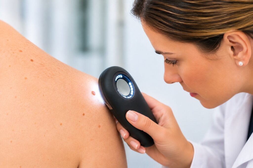
When evaluating suspicious moles and skin lesions, there’s often more to see than what’s on the surface. And while many skin growths are harmless, some may need a closer look. Something that we utilize in our office to do just that is dermoscopy. Dermoscopy is a non-invasive technique to evaluate moles, pigmented lesions, and other suspicious skin growths in great detail.
At Lisa S. Ball, FNP PLLC at the Neiman Dermatology Building in Buffalo, NY, our dermatology providers have completed additional training in dermoscopy, reflecting their commitment to thorough skin examinations, early detection, and high-quality patient care.
What Is Dermoscopy?
Dermoscopy is a non-invasive examination technique that uses a handheld device called a dermatoscope. This instrument provides magnification and specialized lighting, allowing the provider to examine skin structures and patterns that may not be visible to the naked eye.
Dermoscopy is typically used during skin check appointments to help assess:
- New or changing moles
- Suspicious or unusual skin growths
- Pigmented and nonpigmented skin lesions
- Particular features associated with melanoma or other skin cancers
- Lesions that may require further examination or biopsy
By providing a more detailed view of the skin, dermoscopy helps our providers assess skin lesions and determine whether additional evaluation, monitoring, or a skin biopsy is needed.
Why Is Specialized Dermoscopy Training Important?
Dermoscopy is an important tool for evaluating suspicious moles and skin lesions. Using magnification and specialized lighting, it allows providers to identify skin structures and patterns that may not be visible to the naked eye.
Research shows that dermoscopy can improve the accuracy of skin lesion assessments when performed by trained providers.
At Lisa S. Ball, NP PLLC at the Neiman Dermatology Building in Buffalo, NY, all of our dermatology nurse practitioners have completed additional training in dermoscopy to support thorough skin examinations and the early identification of suspicious lesions.
How Does Dermoscopy Help With Skin Cancer Detection?
Dermoscopy can help identify concerning features associated with melanoma, basal cell carcinoma, and squamous cell carcinoma.
During a skin examination, our providers may use dermoscopy to:
- Evaluate suspicious or changing moles
- Identify patterns associated with skin cancer
- Determine whether monitoring or a skin biopsy is appropriate
Dermoscopy is quick, painless, and non-invasive. However, it does not replace a skin biopsy when one is needed to confirm a diagnosis.
When Should You Have a Mole Checked?
The ABCDEs of melanoma can help identify suspicious changes:
- A – Asymmetry: Uneven shape
- B – Border: Irregular or poorly defined edges
- C – Color: Multiple colors or changing pigmentation
- D – Diameter: Larger than 6 mm (size of a pencil eraser), although melanomas can be smaller
- E – Evolving: Changes in size, shape, or appearance
Other warning signs include bleeding, persistent sores, or new or changing skin growths.
Schedule a Skin Cancer Screening in Buffalo, NY
At Lisa S. Ball, NP PLLC at the Neiman Dermatology Building, our dermatology nurse practitioners use their specialized dermoscopy training to evaluate suspicious moles, changing skin lesions, and potential signs of skin cancer.
We provide skin examinations for patients throughout Buffalo, Williamsville, Amherst, Clarence, Orchard Park, East Aurora, and surrounding Western New York communities.
If you’ve noticed a new or changing mole, unusual skin growth, or concerning skin lesion, schedule a professional skin examination for early evaluation.
Schedule Your Skin Examination Online or call 716-688-0020. Learn more about our skin cancer evaluation and treatment services.